BALANCING THE HIND: THE IMPORTANCE OF HOCK JOINT(S)
- Thirza Hendriks

- Jul 10, 2018
- 7 min read

It’s been a while since I posted my last blog about the hip joint in which I promised to follow-up with blogs about the hock and stifle joints. It took me a bit longer as I was just very much investing in field experience, but here it is.
Although the hock is often referred at as if it were a singly entity, it actually is a complex apparatus and might be one of the most regular compromised joints I see in my daily practice. Because the hock has so many interconnections, it is impossible to write about it in an isolated way. Therefore, to understand the hock, we must first understand its function and relations. For the purpose of this post, I’ll elaborate on its Anatomy & Biomechanics in a simplified way.
The hock or ‘tarsus’ is located between the tibia and cannon bone of the horse’s hind limb. It consists of many bones, muscles, tendons, ligaments, blood vessels, nerves, fascia and individual joints (4) that all stabilize and support each other in function.
The top and largest is the Tibio-tarsal joint which is a ‘high motion but low concussion’ joint responsible for 80-90% of the total flexion- extension movement within the hock. The three smaller hock joints in descending order are the proximal inter-tarsal, distal inter-tarsal and Tarso-metatarsal joints. These ‘low motion high concussion’ joints make up for 10-20% of total movement.
I’d like to highlight 3 important functions of the hocks:
- Shock absorption. Because of the horse’s anatomy the hock joints are always under a certain degree of flexion in the early stance phase, enabling it to absorb concussion from the shock waves that directly travel up the limb and the rotational force (torque) produced by the break over phase of the stride.
- Propulsion. In the later part of the stance phase, the hock extends as it generates propulsion to drive the horse forwards.
- Flexing the back. The hock acts in unison with the stifle, but also with the lower back and pelvis. The Sacroiliac (SI) joint connects the sacrum to the pelvis at the ilium. Little movement occurs in the SI joint and it mainly acts as an anchor stabilizing the hind and supporting motion of the Lumbosacral (LS) joint. The LS joint is a hinge between the last lumbar and first sacral vertebrae. In contrast to the SI, the LS allows for a bigger range of motion specialized in flexion/extension, promoting ‘coiling or uncoiling the loins’. The hip joint (see my previous blog) is then needed to engage the hind legs further, allowing even more different directions of movement.
This close interconnection between hip, hock, stifle and lower back is further enhanced by multi-functioning a muscles. For example, Biceps Femoris (hamstrings) extends the hip, hock, stifle and also flexes the latter one. Semitendinosus (hamstrings) extends the hip and hock while flexing the stifle.
It is therefore that a good functioning of the hock is essential in all equine disciplines, yet it is very often compromised. What would be the causes of this?
- Diseases
Probably the most well known are Osteochondrosis (OC) Dissecans (OCD) and Osteoarthritis (OA). These diseases arise as a result of failed vascularization leading to degeneration and is linked to genetics and breeding. However, vascularization could also fail as a result to a bacterial infection (sepsis), physical damage (frostbite) or even trauma. Then, management and nutrition also play a role.
Apart from that many ‘pathologies’ in the hock are described such as bog spavin, bone spavin, blood spavin, throughpin (tarsal tenosynovitis), curb and capped hock (bursitis). However, although these terms imply specific hock conditions, they often are merely a clinical symptom (swelling/ inflammation) of an underlying problem in respectively the bone, tendons (sheath) or ligaments around the hock.
Depending on the location of the swelling the underlying cause can be determined.
X-rays will show changes in joints and bones (OCD and OA) while ultrasound will show changes in soft tissues such as tendons and ligaments.
- Tack
A saddle that is too long could put high pressure on the lumbar area,
affecting the lumbo-sacral joints. As these act in harmony with the hock and stifle, these can get compromised too. Bandages or compression suits could also lead to swellings as it affects the tendons.
- Trauma
Any hard impact such as a kick, getting stuck in the stable or fence could result in fractures, tendon ruptures or a capped hock. Injury to the peroneal nerve leads to alterations in flexor muscles of the tarsus.
- Secondary Trauma
As mentioned before the hock acts in unison with other major joints which could lead to a ‘chicken and the egg’ situation. For example, a horse with OCD in the stifle often starts to rotate at the hip joint to avoid any discomfort. This will also lead to altered movement at the hock. Feet & jaw imbalances, melanoma and shoulder trauma might also lead to hock restriction due to interconnection.
- Breeding
As mentioned before diseases like OCD have a heritable predisposition. Research shows that up to 50% of the Standardbreds and Dutch Warmblood foals have OC lesions. It is a serious problem that gets reproduced year by year. It is worth noting that this disease is almost exclusive to domesticated horses. So it seems that the demand for breeding larger and faster growing horses has ‘accidently’ led to degeneration.
- Confirmation & Natural asymmetry
Every horse has a different confirmation and a preferred side. Some confirmations allow certain disciplines better than others.
- Training
All equine disciplines put a lot of strain on the hocks. Think for example of the spinning and slide stops in western, repetitively going over high fences in show jumping and long periods of collection in dressage.
Too often, a training lacks a ‘proper’ warm-up essential for the joints. I see riders entering an arena with completely loose reins walk around (sometimes even do a quick text or phone call because, hey, we’re just walking right?) a few circles and then pick up the reins and start to seriously trot and then canter. But just walking around isn’t a proper warming up. It is quite the shock both mentally and physically if you just sit on his back and let him walk ‘loose’ and all of a sudden pick up the reins and expect all kinds of (lateral) exercises in balance.
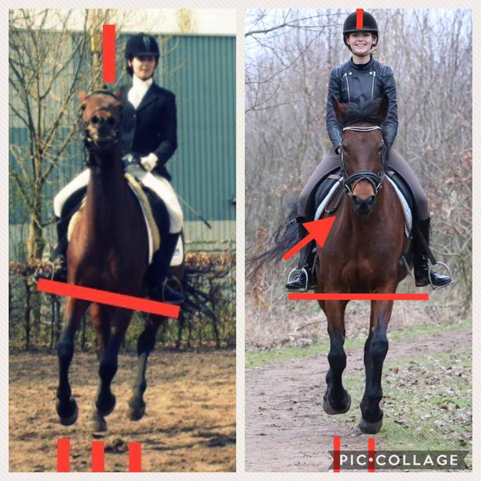
Proper timing of aids is also essential. Too often riders haven’t learned to follow the ‘barrel’ swing properly. The barrel swing tells us when the hind leg takes off into the air to step forward. We can only give an aid during this moment. When we give the aid while the leg is already on the ground, the horse has no option but to start twisting or rotating in the hock and stifle (hip often as well). So we should learn to be still in the movement of the horse. So that we could follow the movement of the horse and not limit or disturb it. Because when we do so, we create an extra shock wave out of the rhythm of the hind legs. Investing in learning a good sitting trot is essential for a healthy (lower) back and hocks!
Also, training in fancy hyperextension , long and low and overflexion will also damage the hocks. The horse often starts to rotate to avoid any discomfort.
The long periods of canter might also contribute to wear and tear on the hocks. In nature, horses often travel long distances in either walk and trot and use the canter only for explosive and short emergency situations to escape predators. However, in modern training there often are required long periods of (collected) canter. In canter, flexion-extension movements are greatest in the lumbosacral region. Research shows linear relationships between LS FE movements and submaximal canter velocities. The required long periods of canter in sport horses puts a lot of strain of the LS joint and therefore the hocks and stifles as well.
Finally, too often we put most stress on what we consider the ‘weak’ parts of our horse. Whenever I meet new combinations, they often tell me whatever is still not going well in their opinion. But what about turning it around? What about considering the strong parts of our self and our horses and start working from that? So if a horse has poor hocks, why trying to put so much focus to make it ‘stronger’ all the time by trying to train up to heavy collection? What about finding what he is really good at, what he really likes and what feels comfortable? To adjust training goals to the different capabitilies of each horse and work from there?
As with all my blogs I can’t give exact tailor-made practical advise on paper but here are some general tips that might help you to ensure a healthy functioning hocks:
- OBSERVE & PALPATE. Look at the confirmation of your horse’s hock. Are they symmetrical? In motion, observe the flexion and extension. Do the hocks rotate in or out? If so, does it come from the hip or is it isolated to just the hocks? How do the hamstrings feel? Are there sign of trauma? Or fibrotic myopathy?
- STOP AT A HIGH. We often spend a lot of time improving things and when it finally goes well, we want to keep going because it finally starts to feel good. But it’s exactly then that we might cause too much wear and tear. So when a horse has weak hocks and all of a sudden gives a really, really nice canter, praise him and stop after a few strides. Because if you keep going on, you risk pushing the limits and spirit of the horse.
- ESSENCE instead of PICTURE. Exercises are meant to be functional, not to look super fancy, which is often a fashion. What is looking fancy now might not be considered fancy in the future. Only with essence in mind can exercises be functional for the horse.
- It’s NEVER JUST TRAINING. It’s always a combination of management, bodywork and (rehabilitative) training. To enhance a good warm-up, you could for example consider to stretch the hind end first, our use balance pads.
So look at your horse. Feel your horse. Ask for help or guidance if you’re struggling. Your horse will be thankful!


Comments